
Ultrasound technology has revolutionized the way we perceive the human body, offering a non-invasive glimpse into our inner workings. Yet, for many, the rainbow of colors displayed on an ultrasound monitor remains a mystery. Among these hues, red and blue are particularly pivotal, often sparking questions about their significance in diagnosing health conditions, including cancer.
Ultrasound imaging, or sonography, employs high-frequency sound waves to produce images of the bodys internal structures. This technique is invaluable for its ability to visualize organs, tissues, and blood flow without the use of radiation. Doppler ultrasound, a specialized form of this technology, adds color to the mix, providing insight into the movement of blood through vessels.
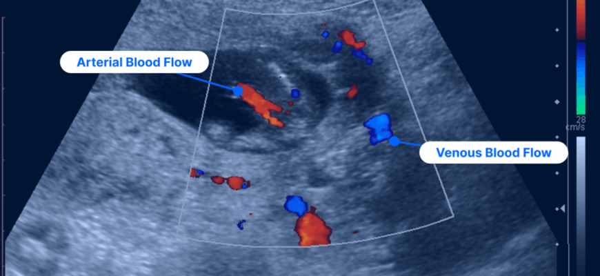
In the realm of Doppler ultrasound, red and blue do not signify danger or safety, but rather, the direction of blood flow. The colors are used to depict the velocity and direction of flow, relative to the ultrasound probe:
- Red: Typically indicates blood moving towards the ultrasound transducer. In clinical practice, this is often displayed as a warm color, suggesting a presence of arterial blood flow.
- Blue: Represents blood moving away from the transducer. This cooler color is commonly associated with venous blood flow.
When it comes to cancer risk, these colors alone do not indicate the presence or absence of cancer. Instead, they provide crucial information about blood flow characteristics that can aid in the diagnosis:
- Increased Blood Flow: Tumors often exhibit increased blood flow, visible as a dynamic mix of red and blue signals. This hypervascularity can suggest malignancy, though it is not definitive on its own.
- Irregular Patterns: The pattern and intensity of the colors, when analyzed alongside other imaging modalities and patient history, can offer clues about the nature of a mass. For instance, chaotic or irregular blood flow patterns may warrant further investigation.
While red and blue can provide valuable insights, they form only a part of the diagnostic puzzle. Radiologists and healthcare professionals integrate Doppler findings with grayscale imaging, patient history, and additional tests such as biopsies or MRIs to arrive at a comprehensive diagnosis.
The interpretation of ultrasound images requires not only sophisticated technology but also the keen eye of an experienced sonographer or radiologist. Their expertise in recognizing subtle variations in blood flow and correlating them with other clinical data is critical in assessing cancer risk.
In essence, the colors on an ultrasound are more than just a visual spectacle; they are a language that, when understood, can reveal vital information about our health. As technology advances, the ability to decode these signals will continue to enhance early detection and treatment of conditions such as cancer, underscoring the importance of informed and skilled interpretation.
Ultimately, while red and blue on an ultrasound can provide meaningful insights into blood flow, they should be considered as part of a broader diagnostic approach. With continued research and technological advancements, their role in the early detection and management of cancer continues to evolve, offering hope for more precise and personalized healthcare.
Ultrasound technology has revolutionized the way we perceive the human body, offering a non-invasive glimpse into our inner workings. Yet, for many, the rainbow of colors displayed on an ultrasound monitor remains a mystery. Among these hues, red and blue are particularly pivotal, often sparking questions about their significance in diagnosing health conditions, including cancer.
- The Basics of Ultrasound Imaging
- Understanding the Color Code: Red and Blue
- Interpreting Colors in the Context of Cancer
- Beyond the Colors: Comprehensive Diagnosis
- The Role of Technology and Expertise
- Conclusion: A Spectrum of Understanding
- The Future of Ultrasound Technology in Cancer Detection
- 3D and 4D Ultrasound: A Leap Forward
- AI and Machine Learning: Enhancing Diagnostic Accuracy
- Empowering Patients Through Knowledge
- Conclusion: A Bright Horizon
The Basics of Ultrasound Imaging
Ultrasound imaging, or sonography, employs high-frequency sound waves to produce images of the bodys internal structures. This technique is invaluable for its ability to visualize organs, tissues, and blood flow without the use of radiation. Doppler ultrasound, a specialized form of this technology, adds color to the mix, providing insight into the movement of blood through vessels.
Understanding the Color Code: Red and Blue
In the realm of Doppler ultrasound, red and blue do not signify danger or safety, but rather, the direction of blood flow. The colors are used to depict the velocity and direction of flow, relative to the ultrasound probe:
- Red: Typically indicates blood moving towards the ultrasound transducer. In clinical practice, this is often displayed as a warm color, suggesting a presence of arterial blood flow.
- Blue: Represents blood moving away from the transducer. This cooler color is commonly associated with venous blood flow.
Interpreting Colors in the Context of Cancer
When it comes to cancer risk, these colors alone do not indicate the presence or absence of cancer. Instead, they provide crucial information about blood flow characteristics that can aid in the diagnosis:
- Increased Blood Flow: Tumors often exhibit increased blood flow, visible as a dynamic mix of red and blue signals. This hypervascularity can suggest malignancy, though it is not definitive on its own.
- Irregular Patterns: The pattern and intensity of the colors, when analyzed alongside other imaging modalities and patient history, can offer clues about the nature of a mass. For instance, chaotic or irregular blood flow patterns may warrant further investigation.
Beyond the Colors: Comprehensive Diagnosis
While red and blue can provide valuable insights, they form only a part of the diagnostic puzzle. Radiologists and healthcare professionals integrate Doppler findings with grayscale imaging, patient history, and additional tests such as biopsies or MRIs to arrive at a comprehensive diagnosis.
The Role of Technology and Expertise
The interpretation of ultrasound images requires not only sophisticated technology but also the keen eye of an experienced sonographer or radiologist. Their expertise in recognizing subtle variations in blood flow and correlating them with other clinical data is critical in assessing cancer risk.
Conclusion: A Spectrum of Understanding
In essence, the colors on an ultrasound are more than just a visual spectacle; they are a language that, when understood, can reveal vital information about our health. As technology advances, the ability to decode these signals will continue to enhance early detection and treatment of conditions such as cancer, underscoring the importance of informed and skilled interpretation.
Ultimately, while red and blue on an ultrasound can provide meaningful insights into blood flow, they should be considered as part of a broader diagnostic approach. With continued research and technological advancements, their role in the early detection and management of cancer continues to evolve, offering hope for more precise and personalized healthcare.
—
The Future of Ultrasound Technology in Cancer Detection
As we venture further into the 21st century, the landscape of medical imaging is rapidly evolving. Innovations in ultrasound technology are paving the way for even more detailed and accurate diagnostic capabilities. From 3D imaging to advanced AI-assisted interpretations, the future holds exciting prospects for enhancing our understanding of ultrasound colors and their implications in cancer diagnostics.
3D and 4D Ultrasound: A Leap Forward
While traditional 2D ultrasound provides a valuable glimpse into the bodys interior, 3D and 4D ultrasound technologies offer a more comprehensive view. By constructing a three-dimensional image, these advanced techniques allow for a detailed examination of anatomical structures, potentially improving the detection of tumors and abnormal blood flow patterns. As these technologies become more accessible, they promise to refine the interpretation of Doppler colors, offering a clearer picture of underlying conditions.
AI and Machine Learning: Enhancing Diagnostic Accuracy
The integration of artificial intelligence and machine learning into ultrasound diagnostics is another game-changer. These technologies can analyze vast amounts of imaging data, identifying patterns and anomalies that might elude the human eye. AI algorithms can potentially recognize subtle changes in blood flow patterns highlighted by Doppler colors, providing earlier and more accurate indications of cancer risk.
Empowering Patients Through Knowledge
In the realm of healthcare, knowledge is power. As patients become more informed about the significance of ultrasound colors and the broader context in which they are interpreted, they can actively participate in their healthcare decisions. By understanding what red and blue truly indicate, patients can engage in meaningful discussions with their healthcare providers, leading to more personalized and effective care plans.
Conclusion: A Bright Horizon
The journey of decoding ultrasound colors is far from over. With ongoing research and technological advancements, the potential to enhance cancer detection and treatment is immense. As we continue to unravel the mysteries of red and blue, we move closer to a future where early diagnosis and intervention become the norm, paving the way for better outcomes and improved quality of life for patients worldwide.
In the end, the colors on an ultrasound screen are more than just diagnostic tools; they are beacons of progress, guiding us toward a brighter and healthier future.
I appreciate how this piece demystifies ultrasound technology. The explanation about how irregular patterns can suggest further investigation is very informative for those curious about diagnostic processes.
I found the explanation about how increased blood flow might indicate malignancy particularly insightful. It’s great to understand that these colors are not about danger but rather about the dynamics of blood flow.
The distinction between arterial and venous blood flow using color was very well explained. This article makes complex medical technology more accessible to the general public.
This was a fascinating read! Understanding that tumors may show increased blood flow as a mix of red and blue signals gives a better perspective on how medical professionals use this information in diagnostics.
I never understood why ultrasounds showed red and blue until now. The article clearly explains their purpose in indicating blood flow direction, which is enlightening for non-medical readers like myself.
This article does an excellent job of explaining the significance of red and blue colors in Doppler ultrasound. It clarifies how these colors relate to blood flow direction, which is crucial for understanding medical imaging.